7450 Slice Chambers
- Designed to be operated as either an interface (Haas type) chamber or as a submerged (Oslo type) chamber.
- Chambers suitable for research into either cell electrophysiology or biochemistry.
- Available in two or four chamber configurations.
- Six-chamber version is available for biochemistry.
- Each chamber is removable from the unit and can be replaced if required.
- Chambers are available manufactured in clear acrylic (Perspex®/Plexiglas®) or PTFE (Teflon®) materials.
Details
This multi-cell slice chamber is designed to work in either Submerged or Interface modes. They features acrylic tissue cell bodies, each with an Ag/AgCl reference electrode for tissue electrophysiology and holding cells designed to keep additional slices viable until used. The unit could of course be used for biochemistry as well. Cell bodies are also available made of Teflon®. This is recommended in settings where pharmacological agents could be absorbed and later released by the acrylic cell body.
Stable, linear flow and temperature are assured in either Submerged or Interface mode. Replacement cell chambers are available in both Acrylic and P.T.F.E. (Teflon®) materials.
A digital display heater / controller unit is included with predictive algorithms (Proportional, Integral and Derivative) and dual feedback from a thermistor for fast feedback, plus another for spot temperature offset. The water bath temperature is controlled to within +/- 0.1 Celsius in a stable environment.
The ergonomics of being able to easily work with multiple slices has been carefully considered in the design of this unit and all multi-chamber systems. Tissue cells are spaced for micromanipulators to hold stimulator and recording electrodes on either side. The 4 channel chambers are the most cost and time efficient. They may be used alone or they can be built up into our semi-automated Workstation or fully automated Synchroslice integrated lab used for hippocampal and heart slice electrophysiology. With optional micromanipulators and overhead cameras, placement of 8 electrodes can be done within 20-30 minutes, with all slices viewed though a quad processor and video monitor.
Chambers for Electrophysiology
- 7450-2A(E) Dual Channel Acrylic
- 7450-4A(E) Quad Channel Acrylic
- 7450-2P(E) Dual Channel PTFE
- 7450-4P(E) Quad Channel PTFE
Chambers for Biochemistry
- 7450-2A(B) Dual Channel Acrylic
- 7450-4A(B) Quad Channel acrylic
- 7450-6A(B) Hex Channel Acrylic
- 7450-2P(B) Dual Channel PTFE
- 7450-4P(B) Quad Channel PTFE
- 7450-6P(B) Hex Channel PTFE
Manual
Last Updated on January 12, 2022
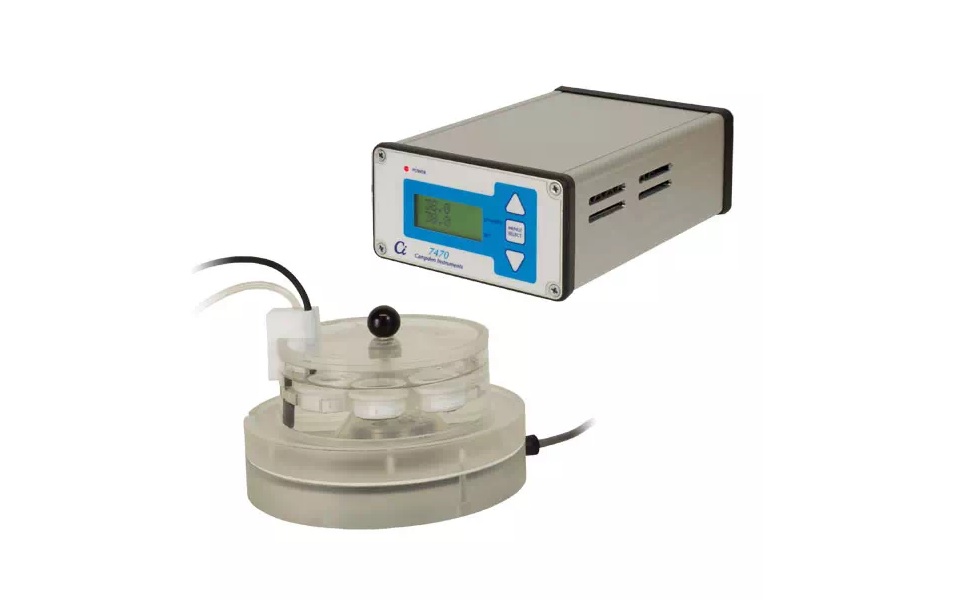
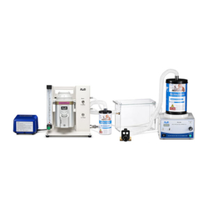
7470 Slice Recovery Chamber
 Six slice holding chambers
- Integrated gas bubbler
- Condensate flow eliminates the risk of osmotic shock
- Bath Thermistor for fast feedback
- Thermistor calibrated to ± 2° C
- Proportional Integral & Derivative Control algorithm
- Overheat Alarm
- Set Temperature Resolution: 0.5°C
- Temperature Resolution: 0.1°C
- Temperature Probe Accuracy: +/- 0.5°C
- Temperature Range: 25°C to 45°C
Details
Simple and Inexpensive!
The Model 7470 Temperature-controlled Slice Holding chamber, has been developed to accompany the Campden Instruments range of tissue slicers.
The 7470 has been developed according to the principles described in the Plymouth Microelectrode Handbook (Gibb, A.J. and Edwards, F.A., 1994. Patch clamp recording from cells in sliced tissues. In Microelectrode Techniques: The Plymouth Workshop Handbook). The chamber was designed to hold and maintain tissue slices in six sub-chambers at physiological temperatures with an aeration supply to allow recovery from slicing as well as incubation during the experimental day (10-12 hrs).
The Proportional Integral & Derivative (PID) controlled heating element is integrated into the stainless steel bath bottom plate. All metal components are manufactured from high-grade stainless steel to remove the risk of contamination of the slice. Temperature feedback is via a thermistor probe attached to the bath wall at the same water as the specimen holder rings. We have also designed the chamber to reduce the risk of thermal/osmotic shock by ensuring the condensation on the lid does not drip back down on to the slice. The aeration pathway has been arranged so that the slice is not disturbed by the bubbles and the created fluid current helps retain the slices within the sub-chamber.
Manual
Last Updated on January 12, 2022
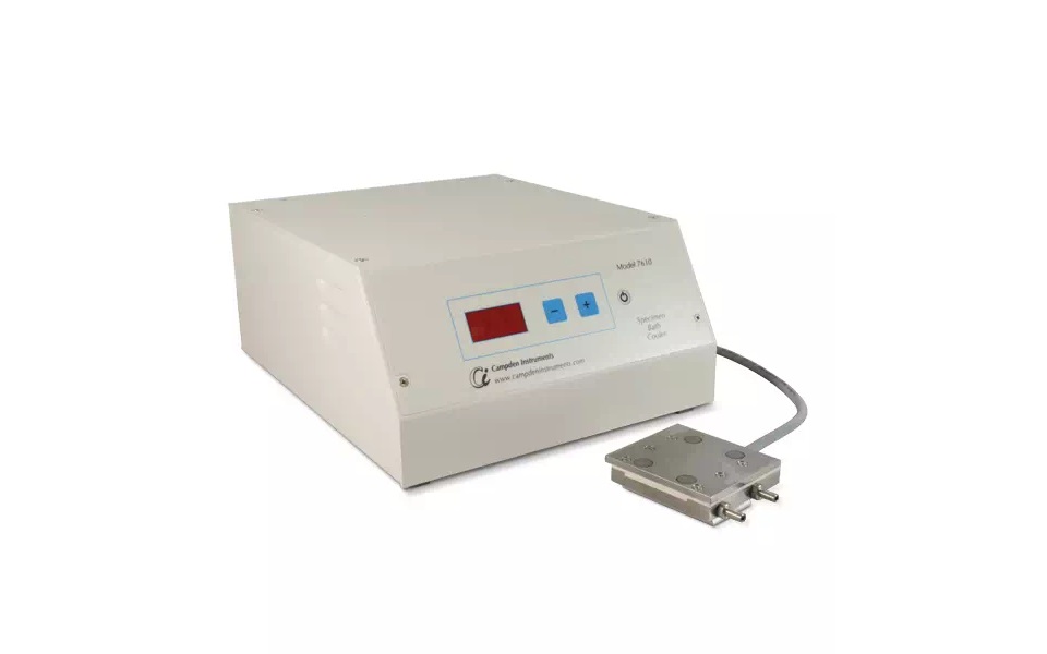
7610A Temperature Controller - Cooling
 Utilitizes the standard inner stainless steel tissue bath
- Display Resolution: 0.1° C
- Temperature Accuracy: +/- 1° C
- Temperature Range: +8-0° C**
- Voltage Requirements: 230V 50Hz or 115V 60Hz
- Power Rating: 60W
- Inlet Fuse Rating: 2A
** Actual temperatures achievable will be dependent upon the solutions used and local temperature conditions. For optimal cooling; inlet water temperature should be below 25° C and flow rate should be greater than 400ml/min
Details
Traditionally once removed from the animal, tissue is immediately cooled to close to +4° C to lower the oxygen demand and prevent anoxia. This has the added advantage with delicate tissues of increasing the stiffness of the tissue allowing for a more robust slice. This can be done with passive cooling by applying ice to the outer icebath (included as standard) or use of the 7610A Tissue Cooler Bath.
The advantage of the 7610A is constant tight control of the bath temperature over long periods without the requirement to refresh the ice or disturb the tissue sample.
The control unit supplies power to the ‘Peltier’ thermoelectric elements in the base of the tissue bath. These act as energy transfer units so that heat is drawn off, through the heat exchanger and removed by tap water flow which runs to waste. The unit uses P.I.D. (proportional integral derivative) temperature control to take the bath temperature to within 0.5° C at the point of measurement. The temperature feedback thermistor is located in the bath floor and software calculates an offset automatically.
Manual
Last Updated on January 12, 2022
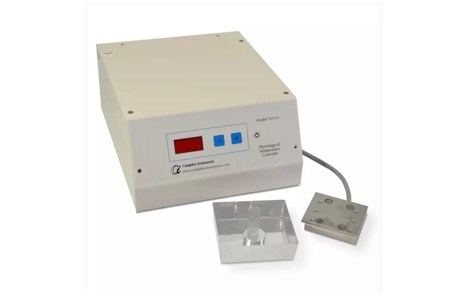
7611A Temperature Controller - Heating
 Utilitizes the standard inner stainless steel tissue bath
- Display Resolution: 0.1° C
- Temperature Accuracy: +/- 1° C
- Temperature Range: +10-50° C**
- Voltage Requirements: 230V 50Hz or 115V 60Hz
- Power Rating: 60W
- Inlet Fuse Rating: 1.25A
** Actual temperatures achievable will be dependent upon the solutions used and local temperature conditions.
Details
Traditionally once removed from the animal, tissue is immediately cooled to close to +4° C to lower the oxygen demand and prevent anoxia. However, we have developed the 7611A due to the paper by Huang S & Uusisaari MY (2013), who demonstrated that slicing at physiological temperature improves the viability and connectivity of the neurons in the slice.
Temperature Control above shown with bath and mount (not included).
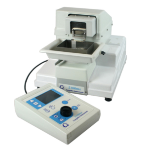
We only recommend the 7611A for use with the 7000smz-2, as this technique has only be shown to be effective if the Z-axis deflection is calibrated to >0.4µm, and the optimal calibrated Z-axis of the 5100mz is 2 ± 0.1µm. The 7000smz-2 Vibrotome has class leading (when correctly calibrated) Z-axis deflection of ≤1 ± 0.1µm at the whole range of speeds and amplitudes of vibration.
The control unit supplies power to the ‘Peltier’ thermoelectric elements in the base of the tissue bath. These act as energy transfer units so that heat is drawn off, through the heat exchanger and removed by tap water flow which runs to waste. The unit uses P.I.D. (proportional integral derivative) temperature control to take the bath temperature to within 0.5° C at the point of measurement. The temperature feedback thermistor is located in the bath floor and software calculates an offset automatically.
Manual
Last Updated on January 12, 2022
7800 Visual Patch & Imaging Chamber
- Easy to use: Just put it on the microscope stage, plug in the controller, connect the tubing and switch it on
- Low profile for easy access of electrodes under the heated objective lens when mounted on upright microscopes.
- No electromagnetic noise emission from the DC power source.
- Annular ring adapter to fit all types of upright and inverted microscopes
- Adapts to 22mm coverslip or 35mm petri dish
- Laminar flow of fluids across the center of the chamber
- 1ml/minutes flow rates
- Used for fever studies at 42° Celsius
- Chambers are coated with chemically inert p.t.f.e. (Teflon®) to avoid adhesion of drugs.
- Gas inlet through heat exchanger into chamber for hypoxia studies, hydrophobic p.t.f.e. resists flooding into gas inlets.
- Unique design of suction capillary ensures a steady state of flow and a mill pond smooth surface in a shallow pool of 1mm depth.
- Ag/AgCl reference wire is simply placed within the chamber
- Vented lid seals the chamber for hypoxia studies
Details
The 7800 Visual Patching and Imaging Chamber consists of an aluminium heat exchanger plate heated by a thin heating element. The heat exchanger supplies concentric heat to the chamber itself and in-line heat to the incoming perfusate ensuring both are heated uniformly. A thermistor embedded in the plate gives temperature feedback to the control system. In its upper surface the plate has a series of annular grooves to carry up to four perfusate tubes. The perfusate tubes carry perfusate to the central chamber at very low level encouraging laminar flow across the chamber.
A gas inlet port allows gas to be introduced for hypoxia studies. Gas flows around the grooves and is heated, eventually exhausting into the central chamber through radial grooves. The heat exchanger plate is coated on all surfaces with p.t.f.e. to discourage drug adhesion. Considerable attention in the design stage has resulted in a chamber with a very low profile reducing the likelihood of the unit interfering with the objective lens and instrumentation probes, electrodes, etc. Because the unit is very compact, the heater output has been closely balanced to the thermal mass of the heat exchanger and the likely demands of perfusate / gas flow. The unit should therefore be protected as much as possible from outside influences such as cold droughts, open doors and windows that may cause unexpected and unwanted variations in temperature.
The temperature controller using PID (Proportional Integral Derivative) algorithms allows close control of the perfusate temperature to within +/- 0.1° C. A second temperature feedback probe can be placed in the central chamber and connected to the controller allowing both chamber temperature and temperature offset (between perfusate and chamber) to be established. The controller has analogue data output capabilities. All power outputs from the controller are DC ensuring that there are no electromagnetic noise emissions to interfere with other instrumentation.
A unique design of suction capillary tube ensures a steady flow of waste perfusate from the centre chamber whilst maintaining a smooth, constant level of perfusate in the centre chamber. The suction tube is adjustable for perfusate depth and has a low profile to avoid interference with other instrumentation probes.
The heat exchanger plate is designed to accept a 22mm slip carrier (along with a vented, double glazed cover if required for hypoxia studies) or a 35mm Petri type dish. Additional slip carriers and chambers to special designs are available on request.
The heat exchanger is mounted on the microscope stage via an insulating adapter ring. The adapter ring has location diameters of 108mm and 110mm making the unit suitable for the majority of upright and inverted microscopes. Other adapter rings to suit a particular microscope can be supplied on request.
Manual
Last Updated on January 12, 2022