Overview
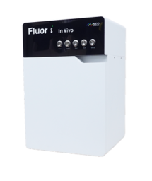
Fluor i In Vivo is a device designed for capturing high-quality images of fluorescent signals in living organisms. It operates using optimized filters that enhances the imaging process by allowing only the emission light to pass through, ensuring clear and detail images. With a fast image processing speed and the innovative Defocusing Free Hyper APO lens, it enables users to capture sharp images across multiple channels without the need for focus adjustments.
Fluor i In Vivo can detect a wide range of fluorescent substances, from blue to NIR, making it versatile for various research applications. Its compact size, along with the separation of luminescent and fluorescent imaging functions, make it easy to use and maintain. This ensures a simplified, yet powerful tool for fluorescence in Vivo imaging.
Last Updated on September 30, 2024
Details
Color Sensor
Flour i In Vivo uses a color sensor to capture and distinguish fluorescent signals from background noise in images. Since fluorescent substances emit different colors depending on their wavelength, a color sensor works better than a black-and-white one for separating the signal from reflected light or auto-fluorescence. This allows Flour i In Vivo to create clearer, more intuitive image data, making it easier to identify the size and location of signals without extra image processing.
Consistent Clarity
Flour I In Vivo’s Hyper Apochromat technology corrects focus for each channel, ensuring consistent focal length across all wavelengths. This results in clearer images and more accurate measurements by eliminating variations caused by light refraction.
Simple
Fluor i In Vivo utilizes a simple, optimized structure, making installation quick and easy. It is also easy to move, manage, and maintain.
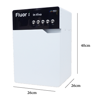
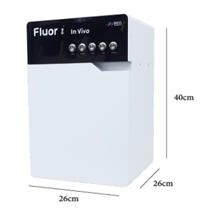
Compact Size
The Fluor i In Vivo has a compact size (26x26x40 cm), so it is ideal for small spaces. Due to its convenient size and portability, it can be used for a wide variety of applications.
Easy to Use
Hardware and software are user-friendly. Live data monitoring, fluorescence intensity comparison, file management, and exposure adjustments are intuitive, with icons and customizable settings, allowing users to navigate effortlessly.
Multi-function
Fluor I In Vivo supports a wide range of applications, from tracking fluorescently labeled drugs to monitoring specific cell types, including tumor, stem, and immune cells. It allows real-time tracking of drug targeting and cell migration, providing precise quantification of fluorescence intensity and area. The device can also be used in plant research to verify gene expression, with specialized filters to remove autofluorescence, ensuring clear imaging of fluorescent genes like GFP. Ex Vivo imaging is available for deeper tissues, offering comprehensive data collection across various experimental setups.
Fig. 3. Animal imaging by Fluor i In Vivo
Last Updated on September 30, 2024
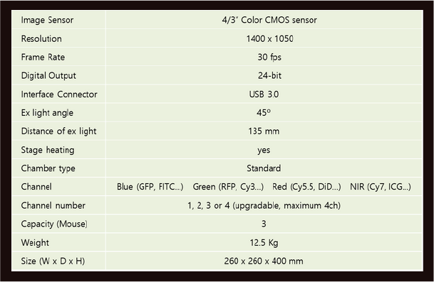
Specifications
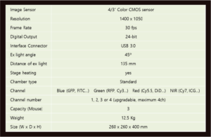
Last Updated on September 30, 2024
Accessories
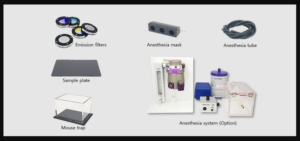
Last Updated on September 30, 2024
Ordering
For ordering information, please contact ALA Scientific Sales team.
Last Updated on September 30, 2024
Most Recent References
- ACS Nano, 2024, Photodynamically Tumor Vessel Destruction Amplified Tumor Targeting of
Nanoparticles for Efficient Chemotherapy.
- Journal of Controlled Release, 2024, Tumor-targeted and stimulus-responsive polymeric prodrug
nanoparticles to enhance the anticancer therapeutic efficacy of doxorubicin.
- Bioactive Materials, 2024, Alveolar macrophage phagocytosis-evading inhaled microgels incorporating
nintedanib-PLGA nanoparticles and pirfenidone-liposomes for improved treatment of pulmonary
fibrosis.
- Acta Pharmaceutica Sinica B, 2024, M1-polarized macrophage-derived cellular nanovesicle-coated
lipid nanoparticles for enhanced cancer treatment through hybridization of gene therapy and cancer
immunotherapy.
- Communications Biology, 2024, The VP53 protein encoded by RNA2 of a fabavirus, broad bean wilt
virus 2, is essential for viral systemic infection.
- Polymers for Advanced Technologies, 2024, Singlet oxygen-responsive hyaluronate dot clusters for
tumor therapy.
- Biomaterials, 2024, Overcoming antibiotic resistance caused by genetic mutations of Helicobacter
pylori with mucin adhesive polymer-based therapeutics.
- Journal of Controlled Release, 2024, Albumin hydrogels for repeated capture of drugs from the
bloodstream and release into the tumor.
- International Journal of Molecular Sciences, 2024, Tumor-Targeted Squaraine Dye for Near-Infrared
Fluorescence-Guided Photodynamic Therapy.
- Materialstoday, 2024, Nitric oxide-assisted mucus-walking zwitterionic nanocomplexes for synergistic
treatment of severe respiratory infectious disease.
- Materials Today Bio, 2024, Amplifying endogenous stem cell migration for in situ bone tissue
formation: Substance P analog and BMP mimetic peptide-loaded click-crosslinked hyaluronic acid
hydrogel.
- Journal of Pharmaceutical Investigation, 2024, Cancer cell membrane-decorated hybrid liposomes for
treating metastatic breast cancer based on enhanced cancer immunotherapy.
- Small, 2024, Protective Topical Dual-Sided Nanofibrous Hemostatic Dressing Using Mussel and Silk
Proteins with Multifunctionality of Hemostasis and Anti-Bacterial Infiltration.
Last Updated on September 30, 2024